
Table of Contents
Context: This year marks the centenary of the first human EEG.
What is Electroencephalography (EEG)?
- Electroencephalography (EEG) is a diagnostic method used to record electrical activity in the brain via electrodes placed on the scalp.
- It is a vital tool for studying brain functions and diagnosing conditions such as epilepsy, monitoring coma and anaesthesia effects, and confirming brain death.
- Invention: EEG was pioneered by Hans Berger in 1924, following earlier research on electrical activity in animal brains by Richard Caton in 1875 and subsequent studies by Adolf Beck and Vladimir Pravdich-Neminsky.
How EEG work?
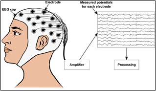
- Principle: Neurons in the brain communicate by transmitting electrically charged particles, creating detectable electrical activity.
- Process: During an EEG test, electrodes capture this activity, which is then analysed to assess various brain functions or diagnose conditions.
EEG Test Procedure
- Electrode Placement: Electrodes are strategically placed using the International 10-20 System, ensuring standardised placement relative to key anatomical landmarks.
- Signal Capture: Neuronal activity causes ions to move, creating waves of electrical activity that are detected by the electrodes and recorded as changes in voltage.
Applications and Limitations
- Clinical Use: EEG is the standard for diagnosing epilepsy and is extensively used to monitor brain activity in various medical situations.
- Research Use: In scientific research, EEG supports studies in neuroscience, cognitive psychology, and other fields.
- Limitations: While EEG is excellent for tracking fast electrical activity, it primarily captures signals from the cortical surface and is less effective at pinpointing the origins of deeper brain activities.
Advantages of EEG
- Cost-Effectiveness: EEG is relatively inexpensive, non-invasive, and portable, making it accessible and convenient for both clinical and research settings.
- Complementary Use: Often used in conjunction with other imaging techniques like MRI to enhance data accuracy and reliability.
Challenges and Considerations
- The setup requires careful preparation, and results can be influenced by factors like the patient’s hair thickness.
- The raw EEG data is affected by volume conduction and must be corrected for accurate interpretation.
- Interpreting EEG data involves understanding complex patterns of voltage and frequency, which require expert analysis.
| Volume Conduction |
| Volume conduction refers to the movement of electrical activity through various media (skin, fluid, bone) before it reaches the electrodes on the scalp. |
 Navy-Marine Expeditionary Ship Interdict...
 K2-18b Exoplanet: Potential Biosignature...
 Vehicle-to-Grid (V2G) Technology and its...